
Our Services
Explore the range of gastroenterology services we offer, along with helpful details and FAQs for many common procedures.
Colonoscopy
Colonoscopy is a relatively painless and usually a quick (30 minutes) procedure. Colonoscopy not only allows for the most thorough evaluation of the colon and identification of polyps and other problems, but also allows for diagnosis, treatment, and removal of potentially precancerous polyps. Colonoscopy is considered the best screening method. It is the only one that allows for the detection, removal and/or biopsy of abnormal tissue as soon as they are detected. A complete bowel cleansing is required before the exam. Colonoscopy is generally done with sedation and is well tolerated. Patients are given medicine through an IV that is placed prior to the start of the procedure to make them feel relaxed and sleepy. Most people do not remember the actual procedure.
-
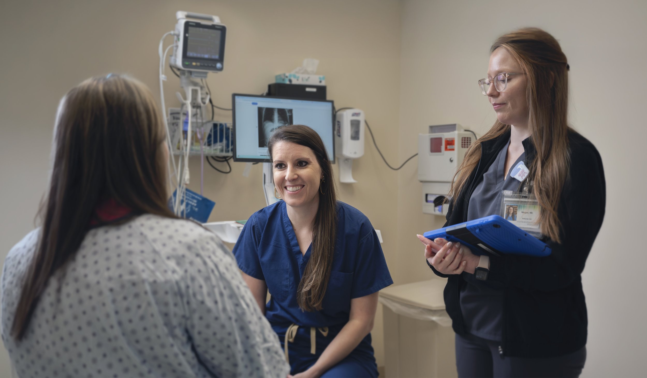
When you arrive the receptionist will call you up to check in. She will verify name, address, date of birth, insurance, etc. You will be asked to sign a few forms.
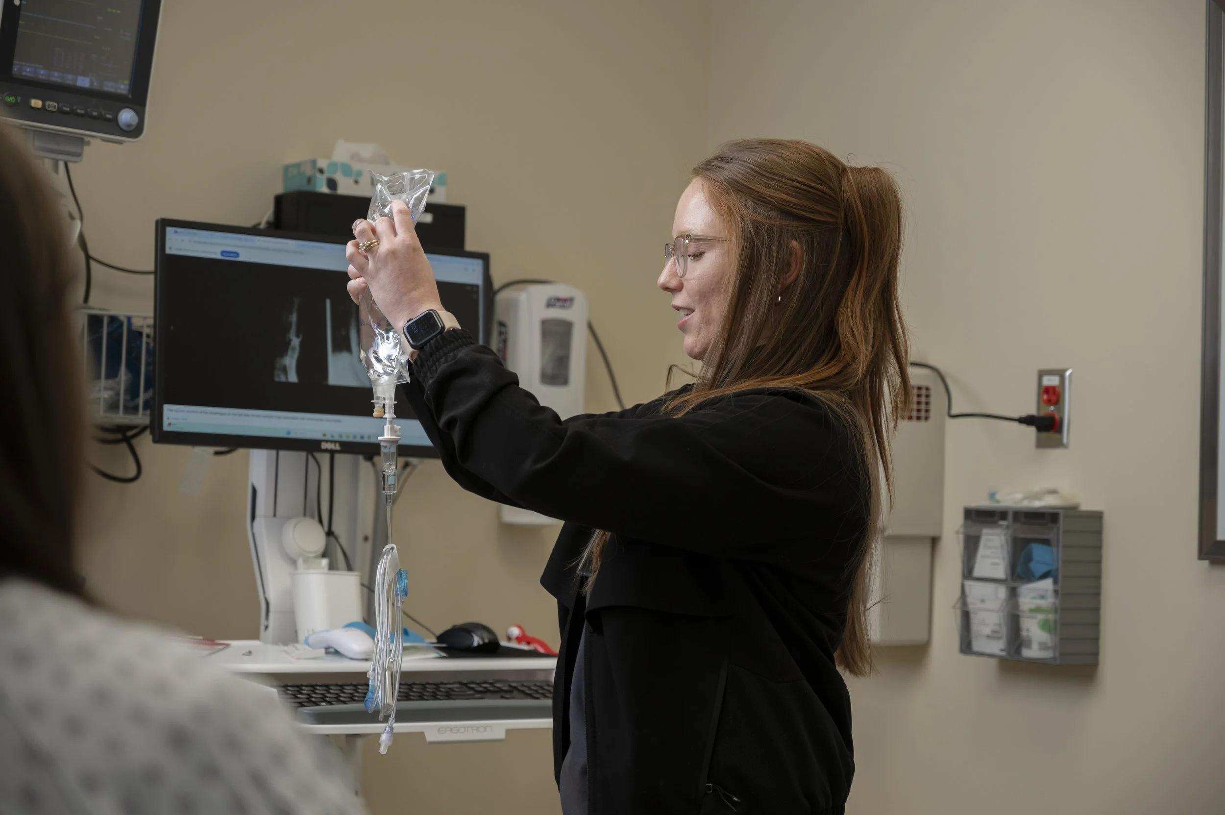
After registration you will be shown to the pre-procedure area where you will change into an examination gown. A nurse will review your medications, allergies and medical history and insert an IV.
Then you will have a chance to speak with your doctor and the anesthesiologist, and proceed to a private procedure suite. You will then be outfitted with several different devices that help monitor your vital signs, etc. during your procedure.
You will be asked to lay on your left side on the bed. You are then given sedation through your IV and will start feeling the effects pretty quickly.
During your procedure if the doctor sees a polyp or other growth or lesion, he/she will remove it. The colon does not feel pinching or burning sensations, so you should not feel polyp (s) being removed.
We recommend that the family member or friend who brought you to the procedure be present when the results are being discussed as you may still be a little foggy from the sedation. Your doctor will also explain any follow up that he/she feels is needed. Patient records are marked for return in three, five or ten years. Our physician will send a letter to you with the results of any tissue sample that were taken within 7-10 days after your procedure.
The nurse will provide you with discharge instructions. Please read over these when you get home. They include things such as diet or medication changes, any follow up instructions from the doctor and an emergency number if you should experience difficulties later in the day. Minor symptoms such as gas or bloating should disappear within 24 hours.
Remember you are not to drive or sign any legal documents after your procedure for the entire day, due to the sedation you received.
-
Anything you can see through such as water, clear fruit juice, carbonated beverages such as ginger ale, clear bouillon, chicken broth, apple juice, white grape juice, plain gelatin Jell-O (no additives), popsicles, coffee, and tea (no creamer and avoid red/purple coloring). Avoid all alcohol the day prior to your procedure.
-
The bowel needs to be cleansed so that the doctor can thoroughly examine the colon. It is important to complete the entire bowel prep; this helps to ensure you will receive the highest quality exam possible. Poor preparation may require a repeat of the colonoscopy on another day so that small polyps (precancerous growths) are not missed due to stool obscuring the exam.
-
There are many types of bowel prep, such as: Golytely, Suprep, Sutab, Suflave, MiraLAX®, etc. Please advise your doctor of any kidney or cardiac problems since some of the preps contain sodium phosphate and may have serious effects on the kidneys. Talk with your doctor about what the best prep might be for you.
-
Possibly. We recommend you contact your insurance company to ask what the preferred medications are on your formulary.
-
The type and severity of side effects differ among patients. They also vary with the product used. Some patients have nausea, vomiting, bloating or abdominal pain and cramping. Read over the instruction packet for more information on the bowel prep that you were given.
-
These are common symptoms that can occur with bowel preparation. Discontinue drinking the prep for 15–60 minutes until resolved and then resume your prep at a slower pace. Drink plenty of other clear liquids. The best prep is the one that is completed; do the best you can.
-
It is not uncommon for your first bowel movement to occur several hours after you begin your prep. Continue to drink plenty of fluids and follow the bowel preparation as advised.
-
We advise that you continue the bowel prep as advised and drink plenty of fluids. We do not suggest taking antidiarrheals as this will counteract the purpose of the bowel preparation.
-
Bring your insurance cards and photo ID.
Bring a responsible adult to drive home (you should not drive the rest of the day).
Bring your payment for deductibles and/or co-payments.
Wear comfortable clothing; elastic waistbands are best. We ask that you not wear jewelry of any type to your appointment.
If you wear a CPAP, please bring it with you. If you have a remote for any implantable stimulators, please bring it with you.
Upper Endoscopy (EGD)
If you have symptoms that do not go away, like heartburn, vomiting or belly pain, you may need an upper endoscopy. An upper endoscopy is also known as an esophagogastroduodenoscopy (EGD). It is a test that enables doctors to examine the upper digestive tract, which includes the:
Esophagus (“food tube” that connects the mouth to the stomach)
Stomach
Duodenum (upper part of the small intestine)
An EGD uses an endoscope, which is a long, flexible tube with a camera and light at its tip. The doctor carefully guides the endoscope through the mouth and down the throat to view the upper digestive tract to view images of the digestive tract and can take color photos of specific areas. They may take a biopsy (tissue sample) of abnormal tissue, such as growths, irritations or ulcers, which are sores in the intestine’s lining.
Flexible Sigmoidoscopy
A sigmoidoscope is a slender, flexible tube with a light and a very small video camera at the end of it. It is shorter than a colonoscope and is only able to evaluate the lower third of the colon. This allows a look at the inside of the rectum and lower part of the colon for cancer or polyps. Before the test, you will need to take an enema or other prep to clean out the lower colon, but a full cleansing solution is not needed as in colonoscopy. This test is often done without any sedation, so it can be uncomfortable, but it should not be painful.
Endoscopic Ultrasonography (EUS)
Endoscopic Ultrasonography (EUS) allows examination of your esophageal and stomach linings as well as the walls of your upper and lower gastrointestinal tract. The upper tract consists of the esophagus, stomach and duodenum; the lower tract includes your colon and rectum. EUS is also used to study other organs that are near the gastrointestinal tract, including the lungs, liver, gall bladder and pancreas. A thin, flexible tube called an endoscope that has a built-in miniature ultrasound probe is used and the endoscope will pass through your mouth or anus to the area to be examined. Your doctor then will use the ultrasound to use sound waves to create visual images of the digestive tract.
Esophageal Dilation
Esophageal dilation is a procedure that allows your doctor to dilate, or stretch, a narrowed area of your esophagus (swallowing tube). Doctors can use various techniques for this procedure while you are sedated during your EGD.
Esophageal Manometry
Esophageal manometry measures the pressures and the pattern of muscle contractions in your esophagus. Abnormalities in the contractions of the esophageal muscle or in the sphincter at the lower end of the esophagus can result in pain, heartburn, and/or difficulty swallowing. Esophageal manometry is used to diagnose the conditions that can cause these symptoms such as achalasia or esophageal spasm.
-
Esophageal testing, also called manometry, measures the pressures and the pattern of muscle contractions in your esophagus. Abnormalities in the contractions and strength of the muscle or in the sphincter at the lower end of the esophagus can result in pain, heartburn, and/or difficulty swallowing. Esophageal manometry is used to diagnose the conditions that can cause these symptoms.
-
An empty stomach allows for the best and safest examination, so do not eat or drink anything for six hours before the test. Since many medications can affect esophageal pressure and the natural muscle contractions required for swallowing, be sure to discuss with your healthcare professional each medication you are taking. Your doctor may ask that you temporarily stop taking one or more medications before your test.
-
A healthcare professional will apply a cream to numb the inside of your nostrils. Then a thin, flexible, lubricated tube will be passed through your nose and advanced into your stomach while you swallow sips of water. Mild, brief gagging may occur while the tube is passed through the throat.
When the tube is in position, you will be sitting upright or lying on your back while the tube is connected to a computer. Once the test begins it is important to breathe slowly and smoothly, remain as quiet as possible, and avoid swallowing unless instructed to do so. As the tube is slowly pulled out of your esophagus, the computer measures and records the pressures in different parts of your esophagus.
During the test, you may experience some discomfort in your nose and/or throat. The test will take approximately 30 minutes to complete and the results will be sent to your doctor’s office.
-
After the test, you may experience a mild sore throat, stuffy nose, or a minor nosebleed; all typically improve within hours. Unless your physician has given you other instructions, you may resume normal meals, activities, and any interrupted medications.
-
As with any medical procedure, there are certain risks. While serious side effects of this procedure are extremely rare, it is possible that you could experience irregular heartbeats, aspiration (when stomach contents flow back into the esophagus and are breathed into the lung), or perforation (a hole in the esophagus). During insertion, the tube may be misdirected into the windpipe before being repositioned. Precautions are taken to prevent such risks, and your physician believes the risks are outweighed by the benefits of this test.
-
In some situations, correct placement of the tube may require passage through the mouth or passing the tube using endoscopy (a procedure that uses a thin, flexible, lighted tube). Your physician will determine the best approach.
Breath Testing
Breath tests are used to identify if your body is malabsorbing specific foods, such as lactose, fructose or sucrose. Breath tests can also be used to diagnose small intestinal bacterial overgrowth (SIBO). Hydrogen breath testing is available in our office.
Hemorrhoid Banding
Hemorrhoid banding, or rubber band ligation (RBL), is a fast and non-surgical approach to hemorrhoid treatment. It doesn’t require fasting, sedation or post-procedure care. And unlike home remedies which provide only temporary relief, it completely and definitively treats hemorrhoid symptoms. It is a simple treatment that can be performed in minutes with little discomfort. No preparation or sedation is needed, and most patients can return to work the same day.
-
Hemorrhoids, also known as piles, are swollen, enlarged veins that appear in the anus or lower rectum. They can be similar to varicose and other varicose veins. Hemorrhoids may develop under the skin around your anus or inside the rectum (internal hemorrhoids).
About three-quarters of adults will experience hemorrhoids at times. There are many causes of hemorrhoids, but the root cause is often unknown.
-
Hemorrhoids refer to swollen blood vessels in the anus. They are quite common in adults and can be very uncomfortable. You can sometimes treat them at home.
Hemorrhoid banding (also known as rubber band ligation) is a method to treat hemorrhoids resistant to home treatment. This minimally invasive procedure involves attaching a rubber band at the base of hemorrhoid to stop blood flow.
-
Hemorrhoid surgery is typically performed as an outpatient procedure. This means that you don’t have to be admitted to a hospital. You might even be able to have it done in your usual office.
You will be anesthetized or have a topical anesthetic applied to your rectum before the procedure. General anesthesia may be required if your hemorrhoids are severe or if you need to have many of them bandaged.
Your doctor will insert an anoscope in your rectum until it reaches your hemorrhoid. An anoscope, a small tube with a light at its end, is what you will see. The anoscope will then insert a small tool known as a ligator.
We use a disposable ligator to create a soft, gentle suction that pulls the appropriate tissue into the device. Then, the rubber band is easily and painlessly placed around the base of the hemorrhoid, where no pain-causing nerve endings are present. This process will be repeated for all other hemorrhoids.
Your doctor will remove any blood clots during the banding process if they find them. Hemorrhoid banding takes only a few minutes in general. However, it can take longer if there are multiple hemorrhoids.
-
Non-surgical hemorrhoid treatment
The procedure only takes a few minutes
No preparation or sedation is needed
Little or no discomfort, virtually Painless!
Most patients return to work the same day
Only for INTERNAL hemorrhoids
-
Tell your doctor about all prescription and over-the-counter medications before the procedure. Also, tell your doctor about herbal supplements.
You may need to avoid eating and drinking for several hours before anesthesia.
Although hemorrhoid banding can be done easily, it is a good idea for someone to take you home and help you around your house for the next day. This will help you avoid straining, which can lead to complications.
Wireless Esophageal pH Monitoring
Wireless esophageal pH monitoring measures the amount of acidic reflux in your esophagus during a 48-hour period and assesses whether your symptoms are correlated with the presence of acid in the esophagus. A small capsule will be attached to the lining of your esophagus to measure and record acidity for 48 hours. As soon as the capsule is attached, it begins measuring the acidity in your esophagus. The capsule sends these measurements wirelessly to a small receiver that you will wear at your waist level held by a strap over your shoulder.
Video Capsule Endoscopy
A procedure that uses a tiny wireless camera to take pictures of your digestive tract. A capsule endoscopy camera sits inside a vitamin-size capsule you swallow. As the capsule travels through your digestive tract, the camera takes thousands of pictures that are transmitted to a recorder you wear on a belt around your waist. Capsule endoscopy helps doctors see inside your small intestine, an area that isn’t easily reached with more-traditional endoscopy procedures.
-
Capsule endoscopy lets your doctor examine the lining of the middle part of your gastrointestinal tract, which includes the three portions of the small intestine (duodenum, jejunum, ileum). Your doctor will give you a pill-sized video camera for you to swallow. This camera has its own light source and takes pictures of your small intestine as it passes through. These pictures are sent to a small recording device you wear on your body. Your doctor will be able to view these pictures at a later time and might be able to provide you with useful information regarding your small intestine.About three-quarters of adults will experience hemorrhoids at times. There are many causes of hemorrhoids, but the root cause is often unknown.
-
Capsule endoscopy helps your doctor evaluate the small intestine. This part of the bowel cannot be reached by traditional upper endoscopy or colonoscopy. The most common reason for doing capsule endoscopy is to search for a cause of bleeding from the small intestine. It may also be useful for detecting polyps, inflammatory bowel disease (Crohn’s disease), ulcers, and tumors of the small intestine. As is the case with most new diagnostic procedures, not all insurance companies are currently reimbursing for this procedure. You may need to check with your own insurance company to ensure that this is a covered benefit.
-
An empty stomach allows for the best and safest examination, so you should have nothing to eat or drink, including water, for approximately twelve hours before the examination. Your doctor will tell you when to start fasting.
Tell your doctor in advance about any medications you take including iron, aspirin, bismuth subsalicylate products, and other over-the-counter medications. You might need to adjust your usual dose prior to the examination.
Discuss any allergies to medications as well as medical conditions, such as swallowing disorders and heart or lung disease. Tell your doctor about the presence of a pacemaker or defibrillator, previous abdominal surgery, or a history of bowel obstructions, inflammatory bowel disease, or adhesions. Your doctor may ask you to do a bowel prep/cleansing prior to the examination.
-
Your doctor will prepare you for the examination by applying a sensor device to your abdomen with adhesive sleeves (similar to tape). The pill-sized capsule endoscope is swallowed and passes naturally through your digestive tract while transmitting video images to a data recorder worn on your belt for approximately eight hours. At the end of the procedure, you will return to the office and the data recorder is removed so that images of your small bowel can be reviewed on a computer screen.
Most patients consider the test comfortable. The capsule endoscope is about the size of a large pill. After ingesting the capsule and until it is excreted, you should not be near an MRI device or schedule an MRI examination.
-
You will be able to drink clear liquids after two hours and eat a light meal after four hours following the capsule ingestion, unless your doctor instructs you otherwise. You will have to avoid vigorous physical activity such as running or jumping during the study. Your doctor generally can tell you the test results within the week following the procedure; however, the results of some tests might take longer.
-
Although complications can occur, they are rare when doctors who are specially trained and experienced in this procedure perform the test. There is potential for the capsule to become stuck at a narrowed spot in the digestive tract, resulting in bowel obstruction. This usually relates to a stricture (narrowing) of the digestive tract from inflammation, prior surgery, or tumor.
It’s important to recognize obstruction early. Signs of obstruction include unusual bloating, abdominal pain, nausea, or vomiting. You should call your doctor immediately for any such concerns. If you develop a fever after the test, have trouble swallowing, or experience chest pain, notify your doctor immediately. Also, be careful not to prematurely disconnect the system as this may result in loss of images being sent to your recording device.
Capsule endoscopy may also be called:
Capsule enteroscopy
Wireless capsule endoscopy
Capsule endoscopy allows for examination of the small intestine, which cannot be easily reached by traditional methods of endoscopy.
PEG Tube Placement & Replacement
PEG stands for percutaneous endoscopic gastrostomy, a procedure in which a flexible feeding tube is placed through the abdominal wall and into the stomach. A PEG tube allows nutrition, fluids and/or medications to be put directly into the stomach, bypassing the mouth and esophagus. A lighted flexible tube called an endoscope will guide the creation of a small opening through the skin of the upper abdomen and directly into the stomach. This procedure allows the doctor to place and secure a feeding tube into the stomach. Patients generally receive an intravenous sedative and local anesthesia, and an antibiotic is given by vein prior to the procedure. You can usually return home the day of the procedure or the next day. Once this tube is in place and can easily be removed at the bedside and if needed, replaced with a new tube once the old tube begins to show signs of wear. This bedside replacement is quick and painless and is performed in the gastroenterologists office.
-
PEG stands for percutaneous endoscopic gastrostomy, a procedure in which a flexible feeding tube is placed through the abdominal wall and into the stomach. PEG allows nutrition, fluids, and/or medications to be put directly into the stomach, bypassing the mouth and esophagus. This brochure will give you a basic understanding of the procedure—how it's performed, how it can help, and what side effects you might experience.
-
Your doctor will use a lighted flexible tube called an endoscope to guide the creation of a small opening through the skin of the upper abdomen and directly into the stomach. This procedure allows the doctor to place and secure a feeding tube into the stomach. Patients generally receive an intravenous sedative and local anesthesia, and an antibiotic is given by vein prior to the procedure. Patients can usually go home the day of the procedure or the next day.
-
Patients who have difficulty swallowing, problems with their appetite, or an inability to take adequate nutrition through the mouth can benefit from this procedure.
-
A dressing will be placed on the PEG site following the procedure. This dressing is usually removed after one or two days. After that, you should clean the site once a day with diluted soap and water and keep the site dry between cleanings. No special dressing or covering is needed.
-
Specialized liquid nutrition, as well as fluids, are given through the PEG tube. If the PEG tube is placed because of swallowing difficulty (e.g., after a stroke), there will still be restrictions on oral intake. Although a few PEG patients may continue to eat or drink after the procedure, this is a very important issue to discuss with your physician.
-
Complications can occur with PEG placement. Possible complications include pain at the PEG site, leakage of stomach contents around the tube site, and dislodgment or malfunction of the tube. Other potential complications include infection of the PEG site, aspiration (inhalation of gastric contents into the lungs), bleeding, and perforation (an unwanted hole in the bowel wall). Your doctor can describe for you symptoms that could indicate a possible complication.
-
PEG tubes can last for months or years. However, because they can break down or become clogged over extended periods of time, they might need to be replaced. Your doctor can easily remove or replace a tube without sedatives or anesthesia, although your doctor might opt to use sedation and endoscopy in some cases. Your doctor will remove the tube using firm traction and will either insert a new tube or let the opening close if no replacement is needed.
Blue Ridge Medical Research
Blue Ridge Medical Research is a division of Gastroenterology Associates of Central Virginia, dedicated to advancing digestive health through clinical trials. We offer patients access to innovative treatments while contributing to the future of GI care.